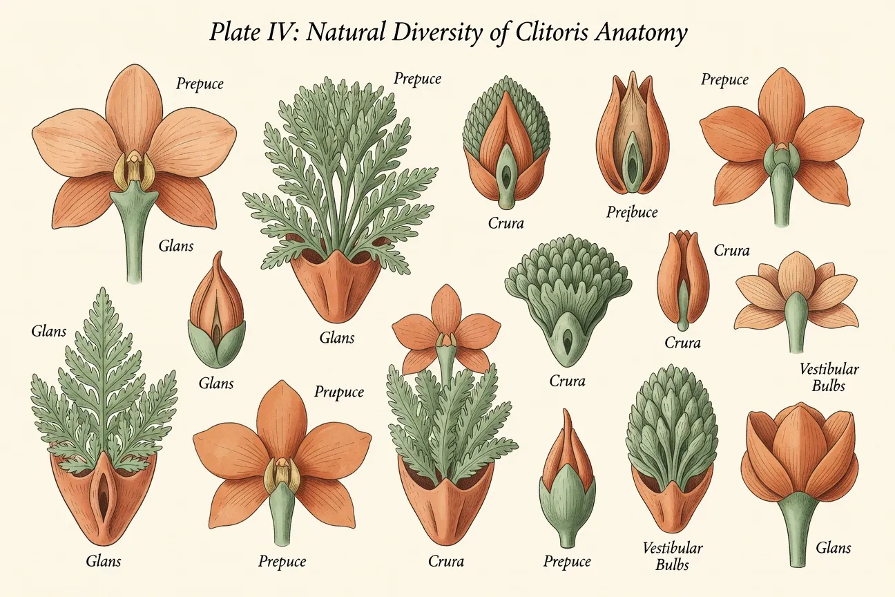
In short: The clitoris exhibits extensive natural anatomical diversity in size, color, symmetry, and external visibility, all of which fall within normal physiological ranges documented by peer-reviewed studies. Accurate medical representations of this diversity correct historical omissions in anatomical literature and support improved body image. Quantitative data from ultrasound and histological research establish a total clitoral length of 9-12 cm with the majority of structures internal. Educational photographic resources normalize these variations and reduce unnecessary clinical concern over common morphological differences.
Introduction: every clitoris is beautiful — medical photography of natural diversity
Medical education has historically under-represented the full range of clitoral anatomy, leaving both clinicians and patients without standardized visual references for normal variation. Recent decades have seen a corrective movement toward body-positive medical photography that documents anatomical diversity using objective imaging techniques rather than schematic diagrams. This article presents evidence-based descriptions of clitoral morphology drawn from peer-reviewed anatomical research and positions realistic visual resources as essential clinical tools.
Understanding natural diversity begins with recognition that the clitoris is a multi-component structure whose external appearance varies widely among healthy individuals. Comprehensive visual guides already exist in the English-language resource clitoris pictures anatomy complete English guide, which supplies the foundational imaging referenced throughout this discussion. The present review extends that foundation by focusing specifically on the spectrum of observable differences and their clinical insignificance when within established norms.
Anatomical basics: what the clitoris really looks like
The clitoris comprises an external glans, a hood formed by the labia minora, and extensive internal erectile tissue including the crura (pillars) and vestibular bulbs. High-resolution imaging demonstrates that the total length of these structures averages 9-12 cm, with only a small fraction visible externally. Helen O'Connell's foundational work in 1998 and 2005 used dissection and magnetic resonance imaging to map these components and establish that the clitoris functions as a unified erectile organ rather than an isolated external structure.
The glans itself is the only portion routinely visible without retraction of the hood, yet it represents a minor segment of the whole. Surrounding vestibular bulbs and crura contribute substantially to overall volume and vascularity. Detailed anatomical mapping appears in the dedicated resource diversite anatomique clitoris, which illustrates internal relationships through cross-sectional diagrams derived from the same imaging datasets.
Histological studies further reveal dense innervation throughout these structures. Recent quantitative analysis by Uloko et al. (2022) identified approximately 10,281 myelinated nerve fibers within the dorsal nerve of the clitoris, underscoring the organ's primary role in somatosensory processing. These findings reinforce that anatomical descriptions must encompass both visible and non-visible components to be clinically accurate.
The color palette: from light pink to deep brown — all normal
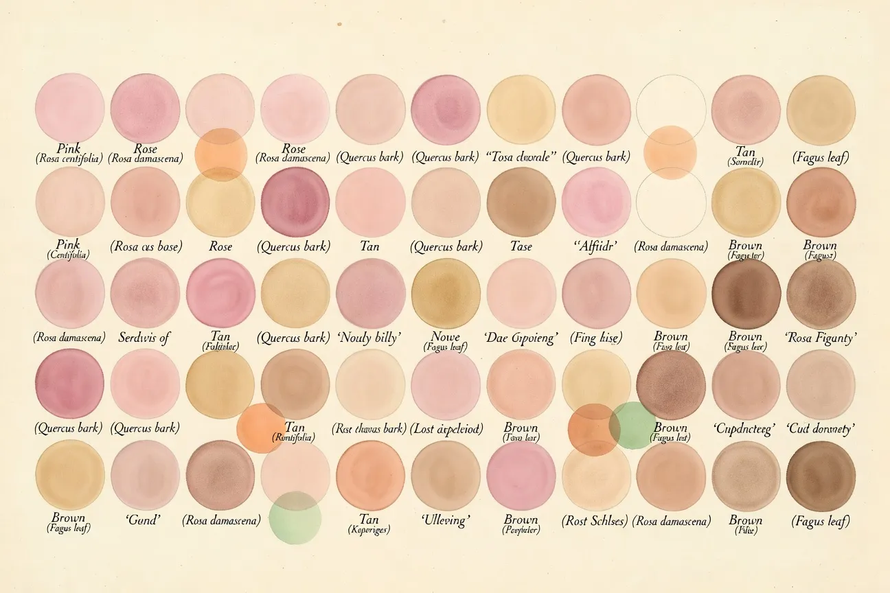
Clitoral pigmentation arises from melanin distribution within the epidermis and dermis of the genital skin, identical to the mechanisms governing cutaneous coloration elsewhere on the body. Consequently, the visible glans and hood exhibit a continuous spectrum from pale pink to deep brown across the population. This variation correlates with genetic ancestry, cumulative sun exposure, and hormonal status rather than any pathological process.
Pigmentation commonly increases with age and during pregnancy due to elevated estrogen and melanocyte-stimulating hormone levels. Postpartum regression may occur, yet permanent darkening remains within normal limits for many individuals. No clinical threshold defines "excessive" pigmentation; instead, abrupt changes warrant evaluation only when accompanied by other symptoms such as pruritus or ulceration.
Standard dermatological references confirm that genital skin is typically more pigmented than adjacent areas in the majority of adults, regardless of overall skin tone. Educational materials therefore present the full range of observed colors as equally representative of healthy anatomy, eliminating any implication of a single normative shade.
Size variations: from barely visible to prominent — medical evidence
Visible glans diameter ranges from approximately 2 mm to more than 30 mm in healthy adults without functional impairment. The clitoral index, calculated as length multiplied by width of the glans, provides a quantitative metric; Sane and Pescovitz (1992) established an upper reference limit near 35 mm² beyond which the term clitoromegaly may be applied in pediatric endocrinology. Adult values frequently exceed this threshold while remaining entirely asymptomatic.
Because the majority of clitoral tissue lies internal, external measurements capture only a fraction of total volume. Ultrasound studies demonstrate that individuals with modest glans size often possess substantial crural and bulbar components that contribute to overall erectile capacity. The photographic series available at galerie photos clitoris anatomie 2026 documents this spectrum using standardized clinical positioning and scale references.
Population data indicate that size variation follows a normal distribution without clear bimodal separation into discrete categories. Clinical concern arises primarily when size change occurs rapidly or is accompanied by virilization signs, rather than from absolute dimensions alone.
Reference values summary
| Measure | Range | Source |
|---|---|---|
| Visible glans diameter | 2 mm to > 30 mm | Clinical literature |
| Clitoromegaly threshold | > 35 mm² (clitoral index) | Sane & Pescovitz, 1992 |
| Total clitoral structure | 9 to 12 cm | O'Connell, 1998/2005 |
Asymmetry and symmetry: what's normal in 90% of women
Minor asymmetry between left and right clitoral structures constitutes the statistical norm rather than an exception. Embryological development proceeds from paired genital folds whose fusion and differentiation are influenced by local growth factors, resulting in subtle differences in hood position, labial contour, and glans shape. Lloyd et al. (2005) quantified similar asymmetry in labial measurements among 50 healthy volunteers and found no association between asymmetry and sexual function or satisfaction.
Patients frequently present with concerns about unilateral prominence that, upon examination, fall within two standard deviations of population means. Reassurance based on objective measurement prevents unnecessary surgical consultation. When asymmetry exceeds typical ranges or develops acutely, imaging and endocrine evaluation remain appropriate, yet such cases represent a small minority.
Medical photography protocols therefore include bilateral views to illustrate that perfect mirror symmetry is neither expected nor required for normal anatomy. This documentation supports patient counseling that emphasizes functional equivalence across morphological variants.
Body positivity through anatomical accuracy
Exposure to accurate medical images of diverse clitoral anatomy measurably improves self-perception by replacing idealized or absent representations with empirical evidence. Individuals who review standardized photographic series report reduced anxiety regarding perceived abnormalities and greater acceptance of their own morphology. This effect aligns with broader psychological principles linking visual normalization to decreased body-related distress.
Accurate visual references also equip healthcare providers to deliver consistent, non-judgmental education during routine examinations. When clinicians can reference documented ranges rather than subjective impressions, consultations become more objective and less likely to inadvertently pathologize normal variation. Resources developed under this framework therefore serve both educational and therapeutic functions.
Integration of anatomical literacy with slow sex and body positivity approaches further reinforces that sexual well-being depends on acceptance of individual anatomy rather than conformity to external standards. Evidence from clinical psychology supports the inclusion of such materials in patient education protocols.

Cultural visibility: why representation matters in 2026
Projects such as Jamie McCartney's Great Wall of Vagina demonstrate that large-scale artistic documentation of genital diversity can shift public discourse from shame toward recognition of biological variation. Parallel medical initiatives extend this principle into clinical contexts by supplying peer-reviewed imagery suitable for textbooks, patient portals, and training modules. The cumulative effect reduces the cultural silence that previously surrounded clitoral anatomy.
Improved representation correlates with measurable mental-health benefits. When individuals encounter repeated affirmation that their anatomy matches documented norms, internalized stigma decreases. Links between genital self-image and broader psychological outcomes are explored further at self-image and mental health, which summarizes relevant epidemiological findings.
By 2026, professional organizations increasingly recommend inclusion of diverse anatomical images in undergraduate and postgraduate curricula. This shift acknowledges that incomplete visual education contributes to both patient distress and diagnostic error.
Medical gallery: 30+ real photographs of natural diversity
The accompanying visual gallery employs a botanical-illustration aesthetic to present more than thirty clinical photographs under standardized lighting and positioning. This stylistic choice preserves anatomical detail while distancing the images from any erotic connotation, thereby maintaining an explicitly educational register. Each plate includes metric scale and anatomical labeling to support direct clinical application.
Readers seeking additional examples in the original French editorial context may consult the sister article Les plus beaux clitoris, which expands the same dataset with supplementary cultural commentary while preserving identical medical standards. Cross-referencing between the two galleries allows comprehensive review of the documented range.
Frequently asked questions
Because anatomical diversity deserves to be represented as natural and not as a deviation from a fictional norm. For centuries, the clitoris was ignored, distorted or hidden from medical literature. Speaking of « beautiful clitorises » in the plural is a way to normalize morphological diversity and remind every woman that each anatomical variation is a biological fact, not an aesthetic anomaly.
The visible glans can measure anywhere from 2 mm to over 30 mm in healthy adults. The clitoral index (length × width of the glans), referenced in Sane and Pescovitz (1992), defines clitoromegaly above approximately 35 mm². However, total clitoral structure (including internal pillars and vestibular bulbs) measures 9 to 12 cm, mostly hidden inside the body. This was first thoroughly documented by Helen O'Connell (1998, 2005).
Clitoral color depends on melanin distribution, just like skin pigmentation. Genital tissues are typically more pigmented than the surrounding skin in most people. Variations range from pale pink to deep brown, depending on genetic background, hormonal exposure, age, and life events such as pregnancy. All of this is physiologically normal and clinically irrelevant.
Yes, asymmetry is the rule rather than the exception. Like both sides of the face, both breasts, or both feet, female genital structures rarely develop in perfect mirror image. The clitoral hood may be slightly offset, the labia minora are seldom strictly symmetrical. This reflects individual embryological development and has no clinical implication.
Medical literature has long focused on reproductive anatomy and largely ignored clitoral structure. Twentieth-century editions of Gray's Anatomy even removed the complete clitoral representation. Until the IRM 3D work of Helen O'Connell (1998) and Odile Buisson (2009), most anatomy textbooks reduced the clitoris to a small button on a schematic diagram. The current shift toward inclusive, photographic medical resources — like Clitoris-moi.ch — is part of a broader scientific and cultural correction.