
In short: This English-language guide gathers carefully labeled clitoris pictures: an external view of the vulva, a sagittal cross-section showing internal anatomy, a frontal view of the legs (crura) and vestibular bulbs, and 3D ultrasounds visualizing the clitoris during arousal. Each illustration comes with a clinical commentary. The guide also documents morphological diversity and the historical erasure of the clitoris from medical textbooks.
Why anatomy pictures matter
For most of the 20th century, the clitoris was almost invisible in medical and educational textbooks. Even today, many school biology books reduce the organ to a small bump on a vulva diagram, ignoring the body, the legs and the bulbs that lie beneath the skin. This long erasure has tangible consequences: complexes, lack of literacy about female sexuality, missed opportunities for clinical care. See also our Beautiful Clitoris: Photographic Medical Guide of Natural Diversity.
Pictures repair what words leave behind. A well-labeled diagram makes it instantly clear that the clitoris extends 8 to 12 cm in total, that its internal portion wraps around the vaginal canal, and that more than 8000 nerve endings concentrate in the visible glans. A single image can replace a thousand awkward conversations.
This English-language guide offers a curated selection of clitoris pictures suitable for adult readers, students, sex educators, doctors and curious learners. Each picture is captioned with a scientific note. The page is designed as a reference: visit, share, bookmark, use in classes.
External view of the vulva and glans
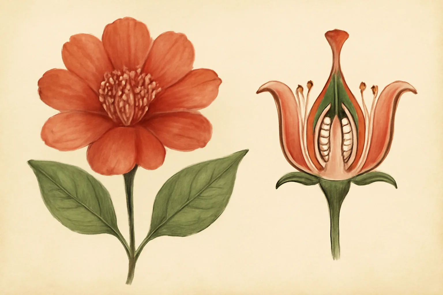
The most familiar image is the external view: a vulva drawn or photographed from below. On such a picture, the visible portion of the clitoris is the glans, a small protrusion located where the inner labia meet at the top of the vulva. It usually sits under a hood, the clitoral prepuce, that protects the highly innervated tissue.
A good external view also labels the mons pubis, the outer labia (labia majora), the inner labia (labia minora), and the vestibule opening to the urethra and the vagina. This view shows roughly ten percent of the clitoral organ. The remaining ninety percent lies internally and requires other types of pictures.
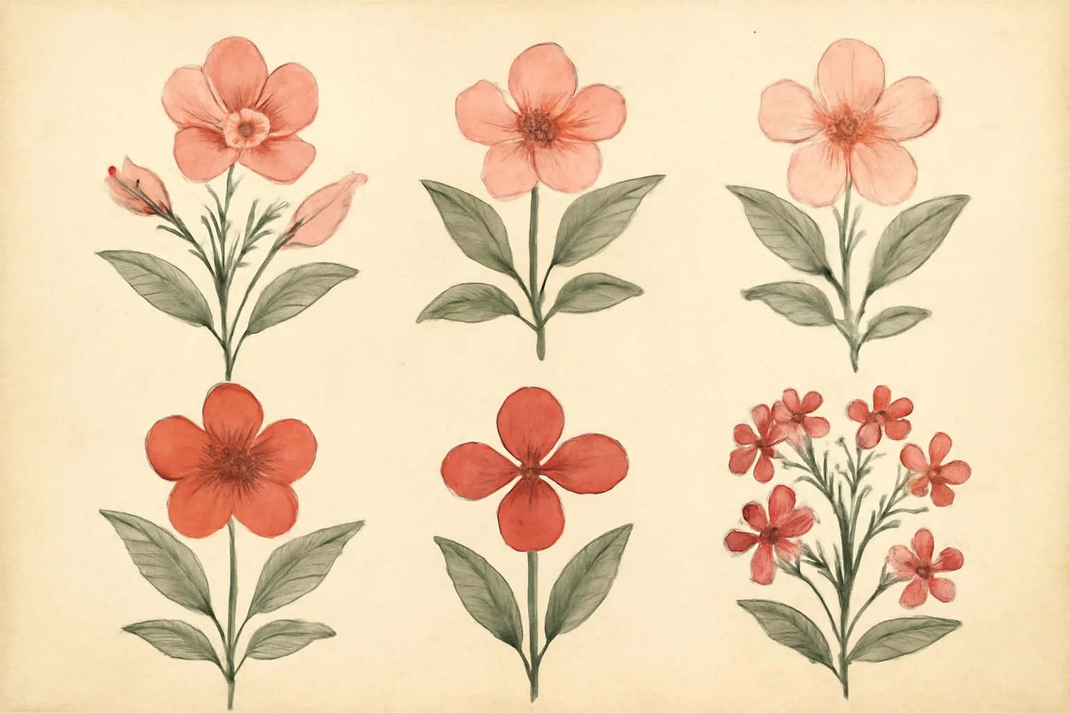
External views also reveal extensive variation: hood length, glans size, labial shape and pigmentation differ widely. For deeper anatomy, see our reference page on the anatomy of the clitoris (in French).
Sagittal cross-section: depth of the clitoris
The sagittal cross-section is arguably the most revealing image. It shows the pelvis cut vertically along its midline, as if the body were transparent and viewed from the side. This picture exposes the body of the clitoris (the shaft) descending into the pelvis, then dividing into two crura (legs) anchored against the pubic bones.
On a sagittal section, you also see the relative position of the clitoris with respect to the urethra, the vagina and the bladder. This explains why both external clitoral stimulation and internal vaginal stimulation can activate the same erectile network: the bulbs and crura lie close to the vaginal walls, and pressure transmits through the tissues. Many older textbooks omitted this view entirely; restoring it is essential for accurate sex education.
Frontal view: crura and vestibular bulbs
The frontal anatomical view shows the clitoris as if lifted out of the body, viewed face-on with surrounding tissue stripped away. Popularized in classroom 3D models since the early 2010s, this image looks like a wishbone or a small flower. From top to bottom, you can identify:
- The glans at the upper apex (3-12 mm in adults).
- The body descending toward the pelvis (2-4 cm long).
- The split into two crura forming an inverted V (5-9 cm each).
- The two vestibular bulbs nestled between the legs, framing the vaginal opening (3-7 cm).
This view dramatically conveys why clitoral sensations can radiate deep into the pelvis. It also illustrates the symmetry and three-dimensional nature of the organ. For an interactive exploration, see our French-language module on the 3D clitoris.
Component dimensions at a glance
| Component | Average size |
|---|---|
| Glans | 3-12 mm |
| Body | 2-4 cm |
| Crura (x2) | 5-9 cm each |
| Vestibular bulbs (x2) | 3-7 cm each |
| Total structure | 8-12 cm |
Comparison with the penis
A complete gallery often pairs a frontal clitoris view with a penile cross-section. The two organs are embryologically homologous: they develop from the same fetal tissue (the genital tubercle), diverging between the 9th and 12th week of gestation under hormonal influence. The glans of the clitoris matches the penile glans; the vestibular bulbs match the corpus spongiosum; the crura match the corpora cavernosa. See our French page on the history of the clitoris for cultural context.
3D ultrasounds: the clitoris in motion
In 2008, French gynecologist Odile Buisson and surgeon Pierre Foldès published the first 3D ultrasounds of the clitoris at rest and during arousal. These images marked a turning point: they confirmed the anatomical hypotheses of Helen O'Connell and showed the organ at work in real time. On a typical clinical ultrasound, the erectile tissue appears as gray spongy areas of varying density.
At rest, the body lies folded against the pubic bone, the bulbs are flat. During arousal, the picture changes visibly: the body swells and elongates, the bulbs nearly double in volume, and the entire complex presses against the vaginal canal. This dynamic view definitively established that the clitoris functions as a complete erectile organ, not unlike the penis.
Ultrasounds are now also used clinically: monitoring tissue integrity after reconstructive surgery (for example, after female genital cutting), evaluating persistent clitoral pain (clitorodynia), or investigating rare congenital variations.
Morphological diversity in pictures
Any responsible educational gallery must show diversity. Pictures depicting only one idealized model reinforce body-image complexes. The reality is that vulvas come in countless variations, all entirely normal. Major variations include:
- Glans size: 3 to 12 mm on average, sometimes larger without underlying pathology.
- Hood shape: short, long, asymmetrical.
- Inner labia length: from a few millimeters to several centimeters beyond the outer labia.
- Pigmentation: pale pink to deep brown, often darker than the surrounding skin.
- Symmetry: most vulvas show slight natural asymmetry.
Artist Jamie McCartney's project Great Wall of Vagina, exhibiting 400 anatomical casts, has rendered this diversity vividly visible. For the medical perspective, see our French-language guide on size and shape of the clitoris.
A short history of clitoris illustrations
Clitoris illustrations have a turbulent history. In the 16th century, Italian anatomists Realdo Colombo and Gabriele Falloppio competed for the « discovery » of the organ, describing it as the « seat of pleasure. » In the 18th century, Caspar Bartholin produced detailed plates of the vestibular bulbs.
The first nearly complete description came in 1844 from Georg Ludwig Kobelt, a German anatomist who depicted the crura, the bulbs and the vascular network with remarkable precision. His plates were almost forgotten in the 20th century, dominated by Freudian theories that hierarchized « mature » (vaginal) and « immature » (clitoral) orgasms and effectively erased the clitoris from textbooks.
It took the work of Australian urologist Helen O'Connell in 1998 to bring the full anatomy back into mainstream science. Using MRI and dissection, she mapped the entire organ. In 2009, Buisson and Foldès added dynamic ultrasound. In 2017, French engineer Odile Fillod created the first open-source, 3D-printable clitoris model, distributed in schools across France and beyond.
Using these pictures in education
An English-language clitoris picture gallery serves multiple audiences. In school sex education, it counters silence and shame by establishing the clitoris as a legitimate organ whose sole function is pleasure. In clinical settings, it helps doctors explain anatomy, surgery or pain to patients. At home, it supports personal exploration and partner communication.
Some best practices:
- Prefer scientifically validated illustrations to anonymous online sketches.
- Combine multiple views (external, sagittal, frontal, ultrasound) to create a 3D understanding.
- Always include diversity to prevent the « single model » trap.
- Explain vocabulary (glans, hood, crura, bulbs) without making it intimidating.
- Connect each image to function: why this structure matters, how it contributes to pleasure or sensation.
For more education resources, see our French-language section on sex education and the clitoris. Mental health is also intertwined with body image; comprehensive resources on the topic can be found at combattreladepression.com (French).
Conclusion
A gallery of clitoris pictures is not a collection of curiosities. It is a piece of restorative education that fills a long, structural silence in medical literature. By showing the organ in its real complexity, depth and diversity, these images give learners the tools to understand a part of human anatomy that has been hidden in plain sight for far too long. Read, share, teach.
Frequently asked questions
Anatomy pictures help learners and patients visualize a structure that words alone cannot fully convey. The clitoris is mostly internal: about 90% of its tissue lies beneath the skin. Diagrams, cross-sections and 3D ultrasounds make the legs (crura), vestibular bulbs and erectile body visible. They reduce shame, dispel myths, and support sex education in schools, clinics and homes.
Externally, the visible part of the clitoris is the glans, a small protrusion at the top of the vulva, usually covered by a hood (clitoral prepuce). The glans averages 5 to 7 mm in length but ranges from 3 to 12 mm in healthy adults. Internally, the clitoris extends as a body, two crura (legs) anchored on the pubic bones, and two vestibular bulbs flanking the vaginal canal. There is no single normal appearance: vulvar morphology varies widely from person to person.
The total clitoris measures roughly 8 to 12 cm including its internal portion, of which only the glans (5 to 7 mm) is externally visible. The body is about 2 to 4 cm long, the crura measure 5 to 9 cm each, and the vestibular bulbs reach 3 to 7 cm in length. These proportions were rigorously documented by Australian urologist Helen O'Connell in 1998.
Yes. In 2008, French gynecologist Odile Buisson and surgeon Pierre Foldès published the first 3D ultrasounds of the clitoris at rest and during arousal. The images visibly show the body lengthening, the bulbs swelling and the entire complex pressing against the vaginal canal. This work confirmed that the clitoris is a fully functional erectile organ, comparable to the penis.
No. Vulvas show wide morphological diversity. The hood may fully cover or barely cover the glans; inner labia may be small, prominent, asymmetrical; pigmentation ranges from pale pink to deep brown. Educational galleries should display this diversity rather than one idealized model. Such variation is the norm, not an exception, and has no impact on sensitivity or pleasure.
Anatomical illustrations and medical pictures are appropriate when they serve education, science and health. They differ fundamentally from pornographic content in framing, intent and accompanying explanation. A scientifically validated clitoris picture, accompanied by anatomical labels and a clear caption, supports literacy about female sexuality. Teachers and clinicians may use such material in non-commercial educational contexts while crediting the source.